☝️ The most important facts in brief
- Nuclear medicine uses radioactive substances to diagnose and treat functional disorders, cancers and other diseases of various organs.
- The type of radioactivity distribution in the body allows doctors to draw conclusions about the underlying disorders.
- To become a nuclear medicine specialist, you must first study medicine and then complete a five-year specialist training programme.
📖 Table of contents
Nowadays, nuclear medicine examinations are essential for the diagnosis of various diseases and functional disorders. Using special radioactive substances that are distributed throughout the body, it is possible to precisely localise centres of disease in organs and tissues. Nuclear medicine is also used for therapy. Find out now how you can become a specialist in nuclear medicine and what exactly you do as a doctor in this speciality.
Are you interested in studying medicine?
We will be happy to advise you free of charge about your options for studying medicine, including advice on studying medicine in another EU country, which is fully recognised in Germany.
What exactly is nuclear medicine?
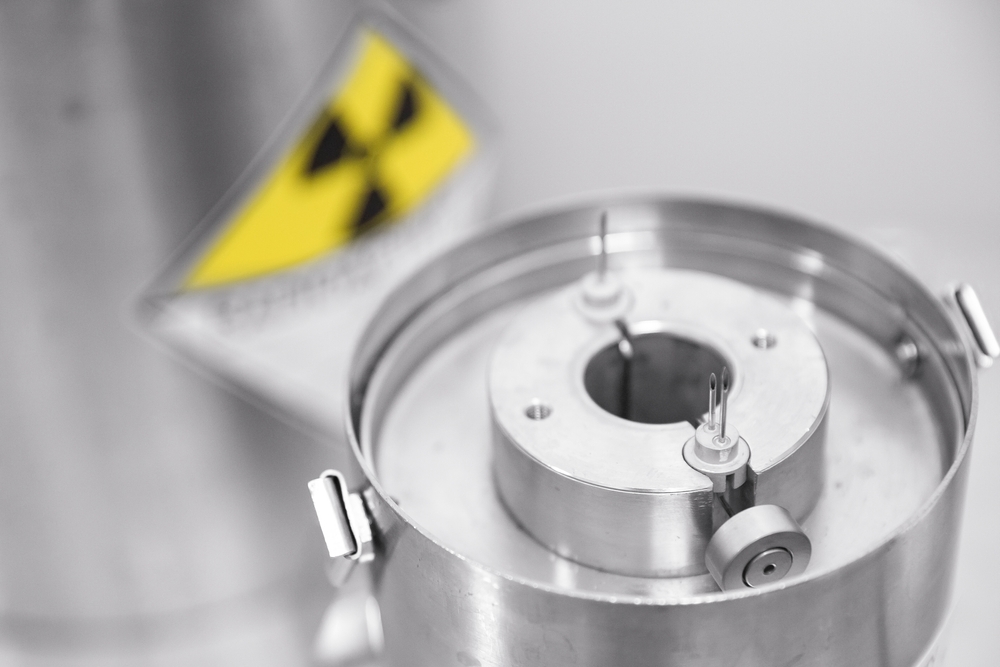
Nuclear medicine is a relatively young medical speciality that uses radioactive substances for diagnosis and treatment. Nuclear medicine examinations can be used to recognise cardiovascular diseases and functional disorders in organs, among other things. So-called radiopharmaceuticals are used for this purpose, which preferentially accumulate in certain cells and thus make it possible to visualise metabolic processes in the body. Nuclear medicine is accompanied by strict radiation protection requirements, but the radiation exposure is generally not too great a risk for the patients concerned.
Tasks of a nuclear medicine specialist
A nuclear medicine specialist can carry out various examinations using radioactive substances and treat patients with special procedures. The examinations in nuclear medicine enable the diagnosis of functional disorders of individual organs and generalised diseases. The core topic of diagnostics in nuclear medicine is above all localisation diagnostics. The radiopharmaceutical specifically selected for the problem at hand reveals the location of the cells that preferentially absorb it. Cancer metastases, for example, can be localised in this way.
Nuclear medicine diagnostics
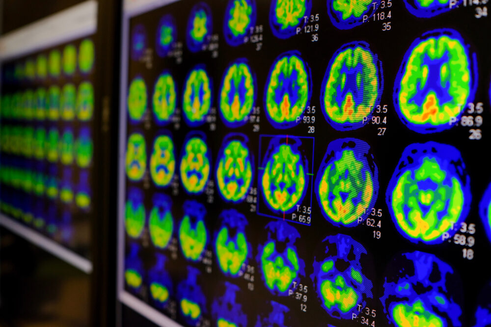
While X-rays, computer tomography or magnetic resonance imaging depict structures of the body, nuclear medicine procedures are used to visualise metabolic processes. By using these examination methods, deviations from the norm can be visualised, which can be caused by cancer or hyperfunction of a certain area, for example. In nuclear medicine, patients benefit from very precise diagnostics with relatively low radiation exposure.
Thyroid scintigraphy and other scintigrams
Scintigrams of the thyroid gland are probably one of the best-known examination and diagnostic procedures in nuclear medicine. Here, the patient is administered the weakly radioactive iodine 131, which accumulates in the thyroid gland. By measuring how much iodine 131 is in which part of the thyroid gland at which time during the examination, the doctor can draw conclusions about the organ's activity.
Scintigrams - with other radioactive substances - are also possible for many other organs such as the heart, kidneys or bones.
Positron emission tomography (PET)
Positron emission tomography (PET) is another nuclear medicine examination that visualises metabolic processes in the body. It also uses a radioactive substance that is absorbed by the tissue and emits so-called positrons, which can be measured. The examination thus provides detailed insights into bodily functions and their disorders.
Radiation exposure and risks from nuclear medicine examinations
Although nuclear medicine examinations involve a certain amount of radiation exposure, they are considered to be very safe for patients. The radioisotopes and medication used are used in small quantities and are quickly excreted.
Nevertheless, safety precautions are important for nuclear medicine examinations and it must be clarified beforehand whether additional risk factors are present.
Nuclear medicine procedures for the treatment of diseases
According to the definition of the discipline, nuclear medicine not only uses scintigraphy and PET scans to diagnose kidney dysfunction, tumours and other diseases, but also carries out treatments. This can often help people for whom treatment with other drugs has not worked.
Tumour cells are irradiated from the inside out
Nuclear medicine can be used to irradiate tumour cells from the inside out. This involves the use of substances that are absorbed by the affected cells due to their specific properties and then emit radiation there. The radiation exposure for the rest of the body is comparatively low.
As a rule, the patient cannot be cured of cancer by these procedures, but the radiation from the nuclear medicine procedures can cause tumours to shrink and may make them easier to operate on.

Relative contraindications
Although the benefits of nuclear medicine examination and treatment methods are generally much greater than the risks, the use of these methods can still be risky under certain conditions.
Particularly in the case of pregnancy or breastfeeding women, it should be carefully considered in advance whether the use of nuclear medicine procedures is appropriate and which protective measures should be observed.
Becoming a nuclear medicine specialist - the contents of the training programme
Training to become a nuclear medicine specialist is extensive and demanding. You must first Study medicine and then undergo further training to become a specialist in nuclear medicine. We would now like to explain these two key stages of training in a little more detail.
Study of medicine
The medical degree programme lays the foundations for future employment in nuclear medicine. You will learn all the important basics about the anatomy and functioning of the body. The various tissues and the details of the metabolism of different organs and their reaction to certain drugs are also part of the degree programme.
Radiology is covered for one semester during medical school, but this course does not yet serve the purposes of specialist training, but is primarily intended to provide an overview for prospective doctors from all specialities.
Specialist training
After studying medicine, you will complete specialist training in nuclear medicine. Among other things, you will learn how to carry out nuclear medicine examinations. You will learn how the metabolism of various organs works in detail and which radioactive substances are therefore best suited to examining these organs.
The training programme includes a lot of theory as well as numerous practical experiences with nuclear medicine examinations and treatment methods. You will complete part of your training in radiology and part directly in nuclear medicine. You can also spend a few months working in a variety of other areas.
Future prospects for the subject of nuclear medicine
Nuclear medicine is still a relatively young speciality in which further progress is constantly being made. Functional and metabolic diagnostics are constantly becoming more precise thanks to new examination methods.
It is to be expected that in future it will be possible to refer patients to nuclear medicine for even more purposes, as new technologies and methods will make this speciality even more effective. Competent nuclear medicine specialists will therefore definitely have good career prospects.
Free information material
Studying medicine abroad 🎉
Order your info pack now, find out more about the Studying medicine abroad and get started as a medical student!


