☝️ The most important facts in brief
- Radiology is a branch of medicine in which imaging techniques are used for diagnostics and radiation and waves are used to treat diseases.
- A radiologist uses technologies such as X-ray, CT and MRI to create detailed images of the body and to be able to recognise bone fractures and other problems, for example.
- The most common activities of a radiologist include the examination of bones, organs and the detection of tumours.
- Further training to become a radiology specialist takes at least 60 months. A medical degree is required beforehand.
📖 Table of contents
Radiology is a fascinating speciality in which an essential part is the diagnosis of diseases using imaging techniques. The radiologist creates images of the body using various technologies. These images help to make the correct diagnosis and initiate the appropriate treatment. In this article, we have compiled a lot of interesting information for you about the work and the various techniques used in radiology.
Are you interested in studying medicine?
We will be happy to advise you free of charge about your options for studying medicine, including advice on studying medicine in another EU country, which is fully recognised in Germany.
What is a radiologist?
A radiologist studied medicine and then underwent 60 months of further training to become a specialist in radiology. During their training, they have familiarised themselves intensively with radiology, computer tomography and magnetic resonance imaging. In addition to imaging procedures, therapeutic interventions are also part of the professional practice of these specialists. For example, radiologists carry out minimally invasive treatments such as embolisation for bleeding and radiofrequency ablation for tumours.
Close collaboration with doctors from other specialities
Both in Germany and in other countries, radiologists work closely with other specialists such as internists and surgeons. They use state-of-the-art equipment and technologies such as X-rays and MRI to ensure the best possible medical care.
Tasks and day-to-day work: What exactly does a radiologist do?
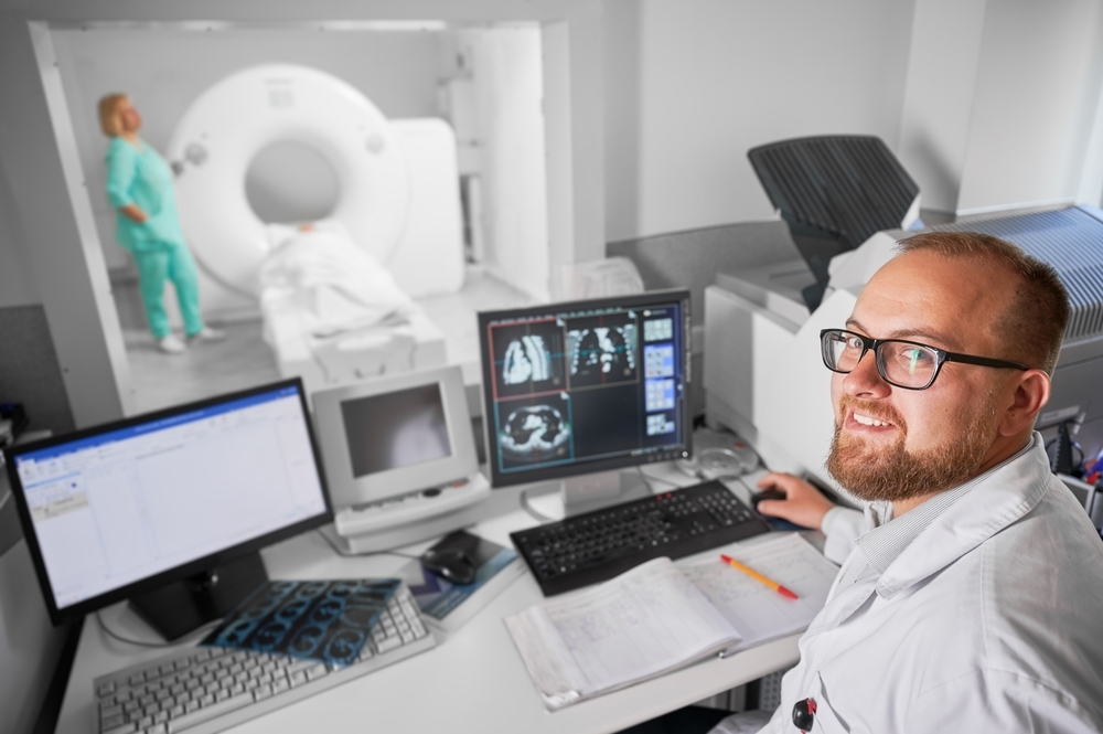
The work of doctors in radiology is extremely varied. They mainly carry out examinations using imaging techniques, analyse them afterwards and discuss the results with patients and doctors from other specialist departments.
Radiologists have various diagnostic procedures at their disposal that fulfil different purposes. You can now get an overview of the most important of these.
X-ray diagnostics
As a radiology specialist, you carry out X-ray examinations to diagnose an existing injury or illness. Thanks to X-rays, you get an accurate picture of the hard structures of the body. Soft tissues such as organs and muscle tissue, on the other hand, cannot be imaged adequately with X-rays.
X-rays are often used for fractures. Bone fractures are visualised in this way so that the doctors treating the patient know after the examination whether a fracture is present and what treatment is appropriate for it.
X-rays are also helpful for diseases of the lungs. Radiation can be used to detect pneumonia or tuberculosis, for example. X-rays penetrate the body. More radiation passes through soft and loose tissue than through hard and solid tissue such as bones. This produces a clear image of the person being treated or the parts of the body being x-rayed, which is not visible from the outside.
Computed tomography (CT)
A radiologist uses computed tomography (CT) to create three-dimensional cross-sectional images of the patient's body. This procedure is particularly valuable in craniocerebral diagnostics and in the diagnosis of strokes.
CT examinations are also important in trauma diagnostics for injured patients. Using X-rays and special equipment, the radiologist creates detailed images that provide information about illnesses and injuries.
CT allows more precise examinations to be carried out than with a simple X-ray. Just like normal X-rays, CT also uses X-rays. The same applies here: The use of this method is particularly useful when viewing bony structures.
Magnetic resonance imaging (MRI)
As we have shown, radiological procedures are a particularly good choice when there are questions about the condition of a patient's bony structures. But what does a radiologist do if there is a suspicion of problems in another tissue and he wants to examine it?
This is where MRI comes in. This procedure does not require X-rays and instead uses magnetic fields and radio waves to produce precise images. This makes MRI examinations a particularly good way of visualising soft tissue such as muscles, ligaments and organs.
Orthopaedic surgeons, oncologists and numerous other doctors and specialists refer their patients to the radiology department in order to check the progression of the disease and gain new insights into the disease by means of an MRI.
Mammography
Mammography is a special X-ray procedure for the early detection of breast cancer. A radiologist uses this procedure to create detailed images of the breast tissue. This enables early diagnosis of breast cancer and increases the chances of successful treatment.
In Germany, mammography is part of the statutory early detection programme for women aged 50 and over and is therefore financed by all health insurance companies. The procedure is quick and usually painless.
By using modern equipment, radiologists can recognise even the smallest changes in breast tissue. Mammography plays a decisive role in the prevention of breast cancer. As with other procedures, however, the images must be analysed with the appropriate expertise. Learning how to analyse such images is part of specialist training in radiology.
Sonography (ultrasound)
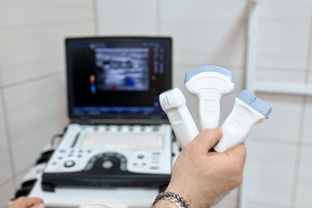
Ultrasound is a particularly gentle diagnostic procedure in radiology. A radiologist uses ultrasound to obtain a two-dimensional image of the body. A 3D ultrasound is now also possible.
The procedure does not involve any harmful radiation and is therefore ideally suited even for pregnant women and children. The ultrasound images provide important information about the organs and tissue structures.
Ultrasound makes it easier than other imaging procedures to observe the processes in the patient's body "live". This makes it easy to check certain functions.
Sonography is used particularly frequently for the examination of abdominal organs, blood vessels and during pregnancy. It is a widely used procedure in Germany.

Training and career as a radiologist
If you decide to specialise in radiology, it will take at least 11 months to get there:
Study of medicine
First of all, you have to complete a medical degree programme. If you are lucky enough to get a place to study medicine straight away, it will take at least 6 years before you can take your state examination and then work as a doctor.
Further training as a radiology specialist
After the state examination, the former students have become doctors who can now take up a position as an assistant doctor at a clinic. To become a radiologist, you still have to complete the 5-year specialist training programme and pass the subsequent examination.
While you are training in radiotherapy, imaging techniques and other areas of radiology, you will be working with patients as a normal doctor. This double burden can be a challenge, but if you are very interested in magnetic resonance imaging, radiology and the like, you will be highly motivated to cope with it and become a qualified radiologist after passing your exams.
What topics are covered in the radiology specialist training programme?
The radiology specialist training programme covers a wide range of topics that you will need for your work later on. The focal points of further training include, among others:
- Imaging procedures such as X-ray, MRI, CT and ultrasound.
- Detailed knowledge of human anatomy and the structures of various tissues.
- The pathology of various diseases
- Radiation protection and safe use of radiation-based therapy and diagnostic procedures
- Interventional radiology procedures
- Dealing with existing diseases and implants that complicate the use of various radiological procedures
Further specialisations are possible within radiology
What does a radiologist do when he has finished his specialist training? - They continue to specialise in a particular area of their field. Of course, the answer to this question is not quite so general, but there are good reasons to actually consider one of the following specialisation options:
Neuroradiology
This speciality focuses on the central nervous system. Among other things, the diagnosis of diseases of the brain and spine using MRI and CT plays an important role.
Paediatric radiology
In many cases, imaging procedures must be adapted to the special needs of children in order to optimise diagnostics for young patients. It is important to use X-rays as carefully as possible and to use the gentlest possible methods for the examinations.
Interventional radiology
This specialist area is dedicated to minimal interventions on the body under imaging control. This can be important when implanting stents, for example.
Career prospects for radiologists
The profession of radiologist is very diverse and it is usually not a big problem to find an attractive position at a clinic. You also have the option of setting up your own practice or working in research and teaching.
What salary can I expect as a radiologist?
Doctors in radiology are among the best-paid specialists in Germany. As a senior physician in radiology, you can expect an average monthly salary of 8,700 euros. If you progress in your career and become a senior consultant, for example, your salary will increase accordingly.
You can earn even more with your own practice. However, this also involves financial risk and costs for equipment and staff.
Differences between radiologists and non-radiologists
Radiology differs significantly from other medical specialisms, such as internal medicine or the Surgery.
Radiologists are specialists in imaging procedures and can also provide valuable services during certain procedures. They have a unique role in large hospitals as well as in private practices. Radiologists work together with many other specialities. If, for example, an MRI is required for a diagnosis, radiology is an important centre of expertise for both oncologists and Sports physician as well as many other specialists.
A radiologist is therefore highly specialised in their field and must also be very familiar with diseases from all other areas. As a radiology specialist, you will see a wide variety of patients and will always be faced with new challenges.
Is radiologist the right profession for you?
The profession of radiologist is not the right choice for every doctor. It combines medical expertise from various fields with state-of-the-art technology. As a radiologist, you will be confronted with a wide variety of tasks and patient fates. You therefore need to have good mental resilience and empathy.
Many of your patients will be afraid of your diagnosis, so it is important to empathise with them and give them bad news appropriately. Depending on your area of work, you will also often have to work under pressure.
Regular further training is mandatory for radiologists
Just as in other areas of medicine, the available options are constantly evolving. As a radiology specialist, it is therefore important to stay up to date. Your training is therefore not over when you take your specialist examination, but extends over the entire time you work as a doctor.
Free information material
Studying medicine abroad 🎉
Order your info pack now, find out more about the Studying medicine abroad and get started as a medical student!


